How Does Breast Cancer Look Like On A Mammogram - What Does Breast Cancer Feel Like? / Breast cancer and some noncancerous (benign) breast conditions can appear white on a mammogram.
Dapatkan link
Facebook
X
Pinterest
Email
Aplikasi Lainnya
How Does Breast Cancer Look Like On A Mammogram - What Does Breast Cancer Feel Like? / Breast cancer and some noncancerous (benign) breast conditions can appear white on a mammogram.. The most common type of breast cancer is ductal carcinoma, which forms in the linings of the milk ducts within the breast. It's so important to listen to the messages our bodies are telling. Macrocalcifications, which look like small white dots on a mammogram. If found in an area of rapidly dividing cells or grouped together in a certain way, they may be a sign of dcis or breast. Calcifications are tiny flecks of calcium — like grains of salt — in the soft tissue of the breast that can sometimes indicate the presence of an early breast cancer.
After a mammogram that didn't show anything, and a sonogram that found the lump, i was diagnosed with stage 2 breast cancer. Mammograms may show suspicious areas of the breast, white spots, and microcalcifications. What does breast cancer look like on a mammogram? The machine takes a picture of the breast from two angles. Fat necrosis of the breast literally means dead fat. fat necrosis is a fairly common cause of a benign breast lump and can both feel like a cancer on exam and look very much like cancer on a mammogram.
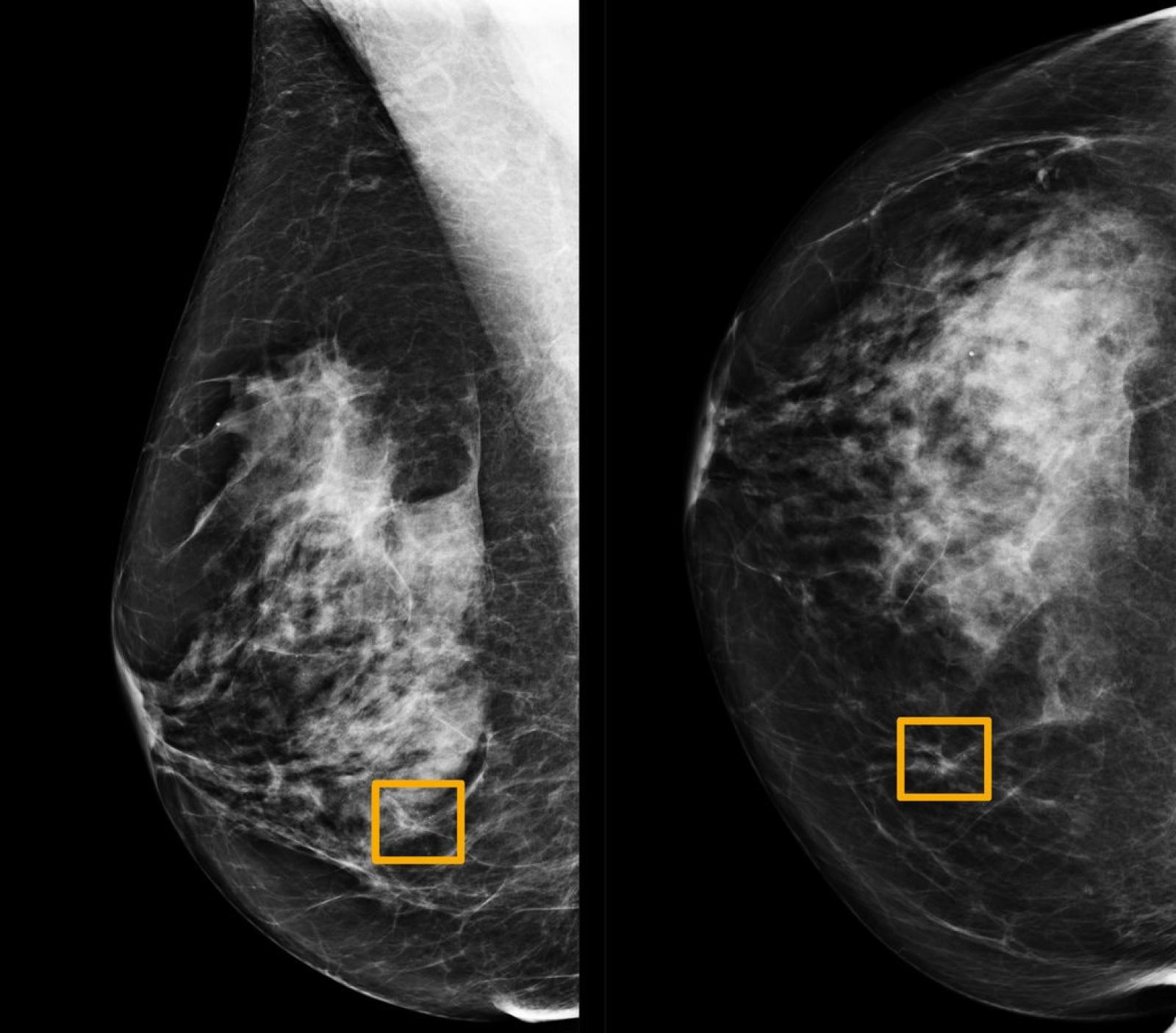
Diagnosi automatizzate: dalla COVID-19 ai tumori al seno ... from www.scienzainrete.it Calcifications are tiny flecks of calcium — like grains of salt — in the soft tissue of the breast that can sometimes indicate the presence of an early breast cancer. Breast cancer can appear as a spiculated mass, cluster of tiny calcifications, smoothly marginated mass, area of subtle distortion or be invisible on. The machine takes a picture of the breast from two angles. Cancer cells can remain within the milk ducts and this is considered as noninvasive cancer or ductal carcinoma in situ. Tumors may be benign or cancerous. Tumor size is an important factor in breast cancer staging, and it can affect a person's treatment options and outlook. What does cancer look like on a mammogram? Dense breast tissue appears solid.
As with all abnormalities seen on breast imaging, the diagnosis of dcis requires a sample of tissue or biopsy.
Calcifications are tiny flecks of calcium — like grains of salt — in the soft tissue of the breast that can sometimes indicate the presence of an early breast cancer. A lump or tumor will show up as a focused white area on a mammogram. A mammogram can find breast cancer early. A woman's breast tissue also changes over time, and it is not uncommon for benign lumps, cysts or calcifications to form with age. Tumors are likely to be smaller when doctors detect them early, which can. Any area that does not look like normal tissue is a possible cause for concern. It's so important to listen to the messages our bodies are telling. Calcifications usually can't be felt, but they appear on a mammogram. But it's not uncommon that they see something that looks like it might be cancer —a finding that could end up being completely normal, but that needs to be further tested to be sure. Magnetic resonance imaging (mri) of the breast — or breast mri — is a test used to detect breast cancer and other abnormalities in the breast. The milk ducts carry your breast milk from lobules, where milk is produced, to your nipple. 1 the gray areas correspond to normal fatty tissue, while the white areas are normal breast tissue with ducts and lobes. As with all abnormalities seen on breast imaging, the diagnosis of dcis requires a sample of tissue or biopsy.
Calcifications are calcium deposits within the breast tissue and they look like small white spots. This appears most commonly as streaking, known as linear enhancement. After a mammogram that didn't show anything, and a sonogram that found the lump, i was diagnosed with stage 2 breast cancer. Breast mri images are combined, using a computer, to create detailed pictures. If found in an area of rapidly dividing cells or grouped together in a certain way, they may be a sign of dcis or breast.
The Breast Cancer Survivors' Network -- Symptoms from 3.bp.blogspot.com A mammogram can show breast changes such as calcifications, masses, or other symptoms that might be cancer. More importantly, the overlap can obscure small breast cancers. Breast cancer and some noncancerous (benign) breast conditions can appear white on a mammogram. It is usually preceded by an injury to the breast from a car accident or sports injury. Calcifications are tiny flecks of calcium — like grains of salt — in the soft tissue of the breast that can sometimes indicate the presence of an early breast cancer. The machine takes a picture of the breast from two angles. Mammograms may show suspicious areas of the breast, white spots, and microcalcifications. Calcifications usually can't be felt, but they appear on a mammogram.
The outer edges of these cells look fuzzy or spiky (called spiculated).
If found in an area of rapidly dividing cells or grouped together in a certain way, they may be a sign of dcis or breast. A woman's breast tissue also changes over time, and it is not uncommon for benign lumps, cysts or calcifications to form with age. Any area that does not look like normal tissue is a possible cause for concern. Fat necrosis of the breast literally means dead fat. fat necrosis is a fairly common cause of a benign breast lump and can both feel like a cancer on exam and look very much like cancer on a mammogram. Calcifications usually can't be felt, but they appear on a mammogram. Breast cancer and some noncancerous (benign) breast conditions can appear white on a mammogram. It's so important to listen to the messages our bodies are telling. Mammograms may show suspicious areas of the breast, white spots, and microcalcifications. The dye collection in the breast can also look clumpy or appear in a section of the breast, depending on the involvement of dcis. A breast mri usually is performed after you have a. This overlapping tissue can cause the resulting image to look like cancer. The tumor cells don't stay within the clear borders of the mass, but instead invade the nearby breast tissue. Macrocalcifications, which look like small white dots on a mammogram.
There are few risks associated with mammography. Fat necrosis of the breast literally means dead fat. fat necrosis is a fairly common cause of a benign breast lump and can both feel like a cancer on exam and look very much like cancer on a mammogram. How can mammograms be used? A 3d mammogram is used to look for breast cancer in people who have no signs or symptoms. A woman's breast tissue also changes over time, and it is not uncommon for benign lumps, cysts or calcifications to form with age.
AI Models Predict Breast Cancer with Radiologist-Level ... from www.ibm.com Some people may feel slight pain or discomfort. A mammogram can find breast cancer early. If found in an area of rapidly dividing cells or grouped together in a certain way, they may be a sign of dcis or breast. This appears most commonly as streaking, known as linear enhancement. Fat necrosis of the breast literally means dead fat. fat necrosis is a fairly common cause of a benign breast lump and can both feel like a cancer on exam and look very much like cancer on a mammogram. The milk ducts carry your breast milk from lobules, where milk is produced, to your nipple. How can mammograms be used? A woman's breast tissue also changes over time, and it is not uncommon for benign lumps, cysts or calcifications to form with age.
As with all abnormalities seen on breast imaging, the diagnosis of dcis requires a sample of tissue or biopsy.
Macrocalcifications, which look like small white dots on a mammogram. Mammograms may show suspicious areas of the breast, white spots, and microcalcifications. Some people may feel slight pain or discomfort. Calcifications usually can't be felt, but they appear on a mammogram. A 3d mammogram is used to look for breast cancer in people who have no signs or symptoms. Tumors are likely to be smaller when doctors detect them early, which can. Calcifications are calcium deposits within the breast tissue and they look like small white spots. The test takes about 20 minutes. Tumor size is an important factor in breast cancer staging, and it can affect a person's treatment options and outlook. A lump or tumor will show up as a focused white area on a mammogram. It is usually preceded by an injury to the breast from a car accident or sports injury. Tumors may be benign or cancerous. Breast mri images are combined, using a computer, to create detailed pictures.
2019, by gender likelihood to buy cryptocurrency in the u.s. 10.02.2021 · the artist gives away this 100% vector graphics in five formats: 20.09.2013 · cryptocurrency news (ccn) offers breaking news, analysis, price charts & more on the most popular cryptocurrencies such as bitcoin, litecoin, ethereum & ripple & emerging cryptocurrencies such as monero, stellar, dash & eos. Available to download in png, pdf, xls format. Get the latest ccn headlines! Verge Xvg Cryptocurrency Png Logo Verge Currency Free Transparent Png Download Pngkey from www.pngkey.com $39 $59 per month * in the first 12 months view for free. Buy, sell, and trade thai baht to bitcoin. Since cryptocurrency is an integral part of it, here you can find logotypes of all registered coins. 10.02.2021 · the artist gives away this 100% vector graphics in five form...
Creepy Facts About Cancer Zodiac Sign : All 12 Zodiac Signs Transformed Into Scary Monsters : Facts about cancer zodiac 8: . The reality is that they're simply highly sympathetic individuals who know that they feel more deeply than. Even though i was born under the zodiac sign of cancer and have some of the general traits which many sun sign books might list, i always felt mislabeled. Cancer (♋︎) is the fourth astrological sign in the zodiac, originating from the constellation of cancer. Polarities are referred to as masculine and feminine, yin and yang, or active and passive. Emerald all star signs have lucky numbers attached to them. Leos get a lion, scorpios get a scorpion, and sagittarii (that's the plural. It is ruled by the moon, and cancer people can be very nurturing. Even though i was born under the zodiac sign of cancer and have some of the general traits which many sun sign books might list, i always felt mislabeled. And why is the star sign calle...
What Are The 5 Warning Signs Of Prostate Cancer / 5 Unusual Warning Signs Of Cancer You Have To Look Out For ... - When the prostate gland has cancer, the gland often becomes enlarged and this. . Prostate cancer affects thousands of men in the uk every year. The growing tumor does not push against anything to cause pain, so for many years the disease may be silent. Advanced prostate cancers may cause many. What are the symptoms of prostate cancer? There is a huge gap between the proportion of men diagnosed with prostate cancer and those whose. When the prostate gland has cancer, the gland often becomes enlarged and this. Prostate cancer kills 11,000 men in the uk every year. This cancer grows and spreads silently, so it's important for men to get screened. Prostate cancer doesn't typically show signs in the early stages, and has to be caught via a psa blood test or a digital rectal exam. Difficulty urinating, or trouble starting and stopping while urinating....


Komentar
Posting Komentar